Syringomyelia Neurology
DISEASE OVERVIEW
Syringomyelia is a clinical syndrome created by a cavity which forms within the spinal cord resulting in the destruction of segmental neurons and long ascending and descending tracts passing through the involved spinal segments. The cavity which forms within the spinal column is called a syrinx (Gr: pipe). A syrinx is cavity is like a pipe or syringe within the gray matter of the spinal column. The two types of syringomyelia are hydromyelia (communicating) and posttraumatic syringomyelia (noncommunicating). The course is usually chronic and progressive.
TYPES
• Posttraumatic: occurs in approximately 3% of patients with spinal cord injury
SIGNS and SYMPTOMS
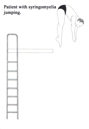
• Symptoms usually begin in the hands and arms (note swimmer diving in hands first)* because of the involvement of the central lower cervical spinal cord.
• Hyporeflexia in upper extremities: due to destruction of the dorsal horn fibers from muscle stretch receptors
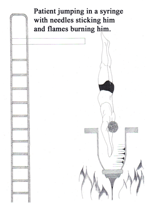
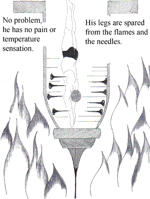
• Loss of pain and temperature perception (hands and arms): destruction of dorsal horn fibers from pain receptors
• Position, vibration, and touch sensation are usually preserved
• Upper extremity muscle weakness and atrophy: destruction of motor neurons and the anterior gray horns
• Lower extremity spastic weakness: occurs as long ascending and descending pathways are compressed by the expanding syrinx
• As the syrinx increases in diameter, long ascending and descending pathways passing through the involved cord segment are compressed or invaded by the cavity and cause spastic weakness of the legs. Tracts to and from the sacral bladder centers are spared, or involved late in the disease, so that bladder symptoms occur late or not at all.
• Central neuropathic pain
• Headache and neck pain
• Autonomic symptoms (interruption of intermediaolateral column): Horner’s syndrome and trophic changes of the skin


DIAGNOSIS
• Spinal MRI with contrast : reveals a central spinal cord cavity
• Electrophysiology: denervation in affected segments
TREATMENT
• Neurosurgical: Budd Chiari malformation – foramen magnum decompression
Posttraumatic - shunt
- Anatomy
- Anesthesiology
- Cardiovascular Medicine
- Dermatology
- Emergency Medicine
- Endocrinology
- Gastroenterology
- Geriatric Medicine
- Gynecology
- HIV
- Hematology and Oncology
- Infectious Disease
- Internal Medicine
- Nephrology
- Neurology
- Obstetrics
- Ophthalmology
- Orthopedics
- Otolaryngology
- Pediatrics
- Pharmacology
- Prevention
- Psychiatry
- Pulmonology
- Radiology
- Rheumatology
- Surgery
- Urology
- case report
- trial reference


